Iron oxide nanoparticles coated with liposomes and chitosan pass a model blood–brain barrier to kill bacteria
(1) St. Michael’s College School, (2) Division of Pre-College and Undergraduate Studies, Brown University
https://doi.org/10.59720/24-056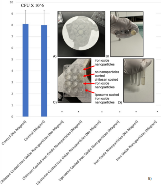
Bacterial meningitis is treated with intravenous antibiotics, which contributes to antibiotic resistance when prescribed widely. The U.S. Centers for Disease Control and Prevention has predicted that one person will die from antibiotic-resistant bacteria every 3 seconds by 2050, representing a significant healthcare crisis. To avoid the use of antibiotics to fight infections, we sought to design, engineer, and characterize iron oxide nanoparticles embedded in liposomes and coated with chitosan to pass the blood–brain barrier to kill bacteria. To create such nanoparticles, the following processes were used: co-precipitation of iron oxide, liposomal encapsulation, and the addition of a chitosan coating. Chitosan was used due to its antibacterial properties. The nanoparticles were characterized by size using dynamic light scattering and tested for their ability to pass through a model blood–brain barrier (filter paper coated with a lipid bilayer) and kill Escherichia coli bacteria. We conducted experiments both in the presence and absence of a magnet to magnetically drive the iron oxide nanoparticles embedded in liposomes and coated with chitosan through a model blood–brain barrier. We observed that the nanoparticles effectively killed bacteria after 12 hours, suggesting that iron oxide nanoparticles embedded in liposomes and coated with chitosan should be further studied for treating bacterial meningitis without resorting to antibiotic use.
This article has been tagged with: